Method Article
Using a 1064-nm Picosecond Neodymium-Doped Yttrium Aluminum Garnet Laser for Periorbital Hyperpigmentation
In This Article
Summary
Here, we present a protocol to describe using a 1064-nm picosecond Neodymium-Doped Yttrium Aluminum Garnet (Nd:YAG) laser with a microlens array for treating periorbital hyperpigmentation.
Abstract
Periorbital hyperpigmentation is a complex condition with multiple underlying causes, such as pigmented, vascular, structural, and mixed factors. The multifaceted nature of the disease presents significant challenges and complexities in its treatment, making it a difficult condition to address effectively. These options include topical cosmetics that can improve the appearance of the affected areas and various chemical treatments. Additionally, fillers are available to enhance volume and smoothen texture, while surgical methods can be employed in more severe cases. Despite these advancements, treating periorbital hyperpigmentation remains challenging. Nowadays, lasers have proven to be highly effective tools in the treatment of a wide range of pigmented diseases. However, there are many types of lasers, and the lack of corresponding guidelines makes the treatment of periorbital hyperpigmentation difficult. Here, we present a protocol describing the use of a 1064-nm picosecond Nd:YAG laser with a microlens array for treating periorbital hyperpigmentation. We discuss optimal energy settings, treatment endpoints, and other side effects while enhancing treatment effectiveness. This approach provides a basis for clinicians to screen and treat patients with eyelids dark circles, ensuring efficacy and safety.
Introduction
Periorbital hyperpigmentation (POH), also known as eye dark circles, is a common cosmetic skin disordercausedby various conditions. Clinically, it presents as symmetric hyperpigmented patches around the eyes distributed on the lower and upper eyelid and may extend to involve the glabella and upper nose1.Some etiologies that contribute to POH are pigmentation, prominent vasculature, skin laxity, and mixed factors.The dark circle around the eyes causes a tired and old appearance, which becomes a psychological concern for patients, so they seek ways to treat it. The disease is easily diagnosed but refractory to treat. Various equipment and tools have been developed to improve the disorder, liketopical cosmetics, chemical peels,lasers, radiofrequency devices, carboxytherapy, fillers, fat injections, and surgical procedures2.
The laser therapy is an effective method for the POH treatment3. Lasers have a selective ability to target endogenous chromophores.The neodymium-doped yttrium aluminum garnet (Nd:YAG) laser is effectively used to treat periorbital dark circles4. The ND: YAG laser is known as a selective photothermolysis system. The laser pulse width is extremely short, which can achieve extremely high peak power in an instant, thereby producing a photoacoustic effect on the target color base. The nonlinear absorption energy of the target color base produces a photodecomposition effect, which ultimately produces a blasting phenomenon leading to the formation of cavitation in the epidermis or dermis, in a process known as laser-induced optical breakdown (LIOB)5,6. There is no damage to the tissues around the LIOB, and the inflammatory reaction is also very slight. With the occurrence of LIOB, new collagen and elastic fibers may appear in the dermis7. The 1064-nm picosecond Nd:YAG laser with fractional microlens array offers several advantages, including effective targeting of pigment particles, a short treatment duration, notable results, and minimal adverse reactions. This article outlines the operation and safety precautions associated with the use of the 1064-nm picosecond Nd:YAG laser for treating periorbital hyperpigmentation and presents clinical cases to illustrate the effectiveness of this treatment.
Protocol
All procedures that involve the participation of human subjects strictly adhere to the established ethical standards set forth by the Ethics Committee of The First Affiliated Hospital of Soochow University and follow the Declaration of Helsinki. Image data collection was conducted with patient consent, and routine examination photographs were captured before treatment.
1. Preoperative evaluation
- Review the patient's medical history.
NOTE: Patient's medical history, including medication history, allergy history, contraindications to treatment, pigmentary changes, previous cosmetic treatments, and manual surgery. - Perform a thorough physical examination. Both the therapist and the patient hold a mirror to examine the area to be treated at the same time.
- Have the patient sign the informed consent form and take the image data using a digital camera and a skin analysis imaging system.
NOTE: Photos were taken at 0°, 45°, and 90° angles before and after treatment, and consistent light and position should be selected when taking pictures.
2. Preparation for laser treatment
- Ask the patient to put on shoe covers when entering the treatment room. Have the patient lie supine to expose the treatment area. Remove jewelry and contact lenses.
- Use a makeup remover and clean the area with a gentle cleaning product.
- Shave the hair in the treated area clean to avoid hair burning and competing with melanin for laser energy absorption.
- Turn on the laser treatment room lights and pre-disinfection of the laser handpiece with 75% alcohol.
- Take a comfortable position, usually sitting, wash hands, and wear a hat, mask, and gloves.
NOTE: Patients use out-of-eye goggles. The therapist wore laser safety goggles for the wavelength used.
3. Treatment
- Choose the Resolve 1064 handpiece (Figure 1A) for the treatment of the patient.
NOTE: The system delivers a 10 × 10 array of 150 µm-diameter microbeams arranged in a 6 mm × 6 mm square treatment area. - Set the energy level between 2.1-2.9 mJ/microbeam, pulse duration of 450 ps, frequency of 5 Hz (Figure 1B).
NOTE: Patients with Fitzpatrick III or IV typically need treatment energy of 2.1-2.3 mJ. - Place the end of the treatment handpiece against the skin, ensuring that the handpiece is perpendicular to the skin.
NOTE: In the periocular area, the orientation of the laser handpiece should be away from the eyeball to reduce the risk of eye injury. - Ensure the pulses overlap by 20% and cover the entire treatment area with laser pulses. Perform the treatment from the side to the middle of the face.
- Aim for the ideal endpoint of treatment, which is the mild darkening of the lesion with slight exudation and bleeding.
NOTE: A 1064 nm picosecond Nd:YAG laser with a mean fluence of 2.1-2.9 J/cm2 was used at 1 month intervals. Treating once a month for a total of 3 treatments.
4. Postoperative care
- Apply an ice pack for 15-20 min after treatment and apply a medium-acting corticosteroid cream twice a day for 3 days.
- Use soothing and moisturizing products for 2 weeks after surgery and avoid harsh skin care products.
- Avoid sun exposure for 4 weeks after treatment to reduce risks such as postinflammatory hyperpigmentation (PIH). Use a broad-spectrum sunscreen with SPF30+ every day, and use umbrellas, hats, goggles, etc.
Results
We evaluated 20 patients aged 21 to 44 years old(8 females and 12 males, mean age 32.4 ± 6.05 years). A total of 4 patients were classified as having Fitzpatrick skin type III, and 16 patients were classified as having Fitzpatrick skin type IV. A total of 7 patients had a family history of POH, and 13 patients had no family history of POH (Table 1).
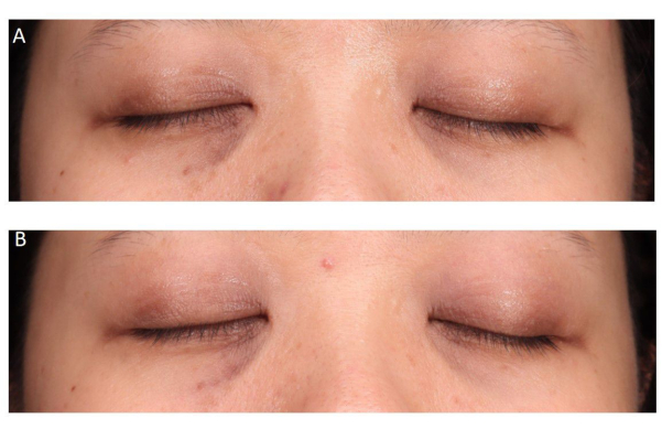
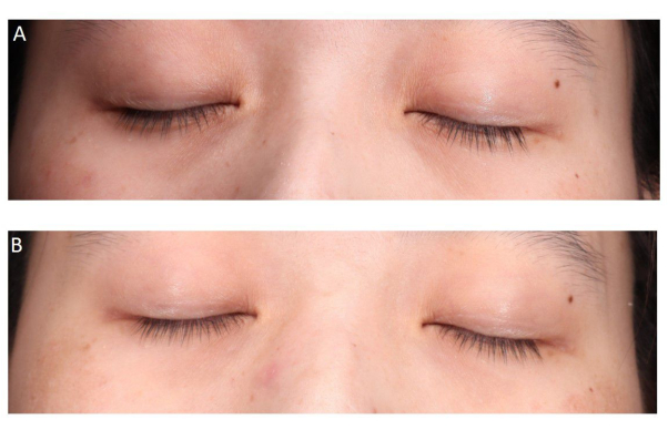
All patients received three consecutive treatment sessions using a 1064-nm picosecond laser with a microlens array at 1-month intervals. The energy was set at 2.3 mJ/µbeam for Fitzpatrick III patients and 2.1 mJ/µbeam for Fitzpatrick IV patients, with a frequency of 5 Hz for all. It is important to cover the entire treatment area with laser pulses during each pass. This process is to be repeated a total of three times to achieve the best possible results. (Figure 2, Figure 3, and Figure 4).
Therapeutic outcome was assessed by two skilled and experienced dermatologists as follows in Table 2. Patient satisfaction was subjectively evaluated after the last treatment using a five-point Likert scale: 1 = extremely unsatisfied, 2 = dissatisfied, 3 = neither satisfied nor unsatisfied, 4 = satisfied, and 5 = very satisfied.
Patients' photographic assessment shows 2 patients had a significant improvement, 11 patients had a moderate improvement, 6 patients had a mild improvement and only 1 patient had no change. Patient subjective assessment revealed that no one was extremely unsatisfied or dissatisfied, 3 (15%) were neither satisfied nor unsatisfied, 12 (60%) were satisfied, 5 (25%) were very satisfied, and the satisfaction scale was 4.1 ± 0.64. The observation found that treatment significantly improved POH compared to baseline (p < 0.05) (Table 3).
The treatment for POH is considered to be a gentle and mild option. None of the patients experienced any serious side effects during the course of the study. Throughout the study, we treated a total of 20 patients, and all of them successfully reached the endpoint of treatment. This endpoint involved mild darkened lesions as well as some slight exudation and bleeding, both of which are expected and temporary reactions. Notably, all of these changes were observed to resolve spontaneously in a few days and did not result in any lingering hyperpigmentation following the treatment. Prior to commencing the laser procedure, we informed all patients about the potential for experiencing pain during the treatment. 12 (60%) patients felt mild pain, 4 (20%) patients felt moderate pain, and 4 (20%) were pain-free. None of the patients chose to withdraw from the treatment process due to the pain, which was within acceptable limits.

Figure 1: 1064-nm Nd:YAG picosecond laser with fractional microlens array. (A) Resolve 1064 handpiece. (B) 1064-nm Nd:YAG picosecond laser parameter adjustment interface. Please click here to view a larger version of this figure.

Figure 2: Frontal views of Case 1. (A) Before treatment and (B) after three treatment sessions. Photographic assessment score: 2. Patient subjective assessment score: 3. Please click here to view a larger version of this figure.

Figure 3: Frontal views of Case 2. (A) Before treatment and (B) after three treatment sessions. Photographic assessment score: 3. Patient subjective assessment score: 4. Please click here to view a larger version of this figure.

Figure 4: Frontal views of Case 3. (A) Before treatment and (B) after three treatment sessions. Photographic assessment score: 3. Patient subjective assessment score: 5. Please click here to view a larger version of this figure.
| Age | 32.4 ± 6.05 (21–44) | |
| Gender | Female | 8 (40%) |
| Male | 12 (60%) | |
| Fitzpatrick skin type | III | 4 (20%) |
| IV | 16 (80%) | |
| Family history | Positive | 7 (35%) |
| Negative | 13 (65%) |
Table 1: Patients' clinical data.
| Score | Progress | Percentage |
| 0 | no change | 0% |
| 1 | mild improvement | 1% - 25% |
| 2 | moderate improvement | 26% -50% |
| 3 | significant improvement | 51% - 75% |
| 4 | excellent improvement | 76% - 100% |
Table 2: Patients' photographic assessment score.
| Photographic assessment | |
| No change | 1 (5%) |
| Mild improvement | 6 (30%) |
| Moderate improvement | 11 (55%) |
| Significant improvement | 2 (10%) |
| Excellent improvement | 0 (0%) |
| Patient subjective assessment | |
| 1 (Extremely unsatisfied) | 0 (0%) |
| 2 (Dissatisfied) | 0 (0%) |
| 3 (Neither satisfied nor unsatisfied) | 3 (15%) |
| 4 (Satisfied) | 12 (60%) |
| 5 (Very satisfied) | 5 (25%) |
| Likert satisfaction scale | 4.1± 0.64 |
Table 3: Treatment outcome of the patients by photographic and subjective assessments.
Discussion
POH manifested as brown or dark brown pigmentation spots in the bilateral periocular area. There are different causes of periorbital hyperpigmentation, such as genetics, gender, age, physical conditions, anatomical differences, excessive blood vessels, etc. It is a concern at any age because it can make a person look sad, old, or tired and weaken their sense of well-being and self-esteem. The therapeutic approach must vary depending on the cause, as POH is caused by a variety of factors. For POH due to melanin, there are currently different treatment options, such as topical medication, chemical stripping, and laser. For POH caused by the thin eyelid skin on the orbicularis oculi muscle, the treatment is to restore the volume under the eyelid, such as fillers and autologous fat transplantation8,9,10.
Lasers have been widely used in the treatment of POH, such as Q-switched, long-pulsed, fractional CO2. An important principle of laser therapy for POH is its ability to selectively target endogenous chromophores such as melanin and hemoglobin. Melanin has a broad absorption spectrum, with the strongest absorption in the ultraviolet range and decreasing gradually with increasing wavelengths. Due to the short thermal relaxation time of melanosomes (<1 ms), an extremely short pulse duration is required to limit the photothermal and photoacoustic effects of melanin11. In recent years, low-flux Q-switched Nd:YAG lasers (LFQSNY) have been widely used to treat melasma, especially in Asia, commonly referred to as 'laser toning (LT)'. LT is known to be delivered multiple times through large spot sizes to optimize energy transfer12. In the treatment of POH as demonstrated in the treatment of melasma, repeated use of LFQSNY (higher fluences than for melasma) treatment can reduce the amount of melanin and thus increase the brightness of the skin. The 1064-nm picosecond Nd:YAG laser delivers energy in nanosecond pulse durations and is capable of targeting melanin. Using the principle of photoacoustic action, the laser instantly produces high energy and is absorbed by the pigment particles to expand and rupture, thereby shattering the melanin. Then, the subcutaneous melanin is decomposed into small and easily metabolized particles, similar to dust13. The 1064-nm Nd:YAG picosecond laser with fractional microlens array emits many beams of small and uniform diameter and uses the principle of fractional photothermolysis to create microscopic columns of thermal injury in the dermis, called microthermal zones (MTZs). The laser eliminates melanin pigment from the epidermis and dermis through a "melanin shuttle" which exudes the pigment from the skin through the MTZs14.
1064-nm picosecond Nd:YAG laser with microlens array helps to promote the production of new collagen, elastic fibers, and growth factors and improves skin texture. This treatment is frequently employed to enhance the appearance of enlarged pores as well as to help in the improvement of scars, which can include post-acne scars, atrophic scars, and posttraumatic and postsurgical atrophic scars15,16,17. The application of this treatment for managing various pigmented diseases is seldom documented in the available literature. This study is limited by its focus on a single ethnicity, lack of diversity in Fitzpatrick skin types, and a small sample size. It is essential to conduct future studies that are prospective in nature and encompass a large-scale population comprising various ethnicities as well as different Fitzpatrick skin types. Additionally, it is important to recognize that adverse events or side effects that may arise following laser treatment could vary significantly among different individuals, highlighting the need for a more extensive understanding of these differences.
The significance of the method with respect to existing methods is as follows. Firstly, the extremely short pulse width of the picosecond makes it more suitable for blasting pigment particles with smaller diameters, while the pigment particles in POH are smaller than those in diseases such as freckles. Secondly, the 1064 nm band has a selective photothermal effect on blood vessels, and the formation of POH includes prominent vasculature, which is more suitable and effective for POH patients. Finally, the skin around the eye is very thin and tender. If the use of ordinary Q-switched or picoseconds, then it is easy to cause secondary color sinking or color loss, causing the local situation to become worse. The fractional mode has lower energy than a single beam, but at the same time retains the effect of selective photothermal effect, which can be relatively good to improve POH.
Some issues that should be noted throughout the laser treatment as listed below. Take a detailed medical history to determine if there are activities that interfere with treatment, such as prolonged daylight exposure, outdoor surfing, swimming, etc. Treatment contraindications include active infection in the treated area, skin diseases in the treatment area such as vitiligo, psoriasis, atopic dermatitis, the use of tanning products 2 weeks before treatment, the use of photosensitive drugs, photosensitive diseases, pregnancy or lactation, the patients' unrealistic expectations.
Determine whether the skin lesions to be treated are complicated by telangiectasia, skin relaxation, wrinkles, and other skin diseases. If blood vessels are abundant, vascular dilation can be treated first. If the blood vessel dilation is not visible to the naked eye, the 1064 nm laser can also partially improve it. The combination of skin relaxation and wrinkles often indicates that the patient's skin photodamage is excessive. The arrangement of elastic fibers and collagen fibers in the skin is disordered, and the shadow around the eyes may be deepened due to local stereoscopic contour changes in such patients. The laser treatment is not effective, and surgical treatment is required.
Patients seeking cosmetic treatments have higher expectations and a lower tolerance for side effects. Before treatment: a detailed discussion of the efficacy and side effects, alternative treatment methods, and their advantages and disadvantages. Give the patient enough time to ask questions and resolve doubts. Inform the patient of the nature and details of the treatment program.
Laser treatment of periorbital hyperpigmentation does not require conventional anesthesia.Anesthesia may interfere with patient response and affect the choice of laser parameters.If the patient has a low pain threshold, nonsteroidal anti-inflammatory drugs (NSAIDS) analgesics can be taken orally 1 h before treatment.
The energy density was selected based on the Fitzpatrick skin type of the patient and the characteristics of pigmented lesions in the treatment area, and the frequency was selected based on the size of the treatment area. When using high-frequency therapy, keep the pulses adjacent to each other to avoid missing the treatment area. A higher energy density can be used for light-skinned Fitzpatrick skin type patients, and a lower energy density can be used for dark-skinned Fitzpatrick skin type patients.The laser-tissue interaction and treatment endpoint were closely observed during treatment. If an adequate treatment endpoint is not achieved in scattered lesions, the treatment endpoint may be enhanced with the same or slightly lower energy density.
Mild edema, erythema, and purpura are normal and usually resolve spontaneously within a few hours to a few days after treatment. Mild itching may occur in the treated area, which usually resolves spontaneously in 3 days without special treatment. Pigmented lesions become darker about 1 day after treatment, indicating the formation of micro-scabs, but they cannot be touched. The scab will peel off about 1 week after treatment, leaving a shallow skin lesion.
For dark-skinned Fitzpatrick skin type patients, kojic acid, arbutin, niacinamide, and other topical products can be applied 1 month before treatment to reduce the pigment synthesis and the side effects of post-inflammatory hyperpigmentation. It is crucial to fully understand the patient's skin condition and the equipment's condition and select the appropriate treatment parameters. For different indications, the next treatment should be performed according to the interval cycle prescribed by the doctor.
Disclosures
The authors have no conflicts of interest to declare.
Acknowledgements
None.
Materials
| Name | Company | Catalog Number | Comments |
| 1064-nm Nd:YAG picosecond laser | Syneron-Candela, Wayland, MA, USA | Resolve 1064 handpiece | 1064-nm Nd:YAG picosecond laser with fractional microlens array |
References
- Roberts, W. E. Periorbital hyperpigmentation: review of etiology, medical evaluation, and aesthetic treatment. J Drugs Dermatol. 13 (4), 472-482 (2014).
- Michelle, L., Pouldar Foulad, D., Ekelem, C., Saedi, N., Mesinkovska, N. A. Treatments of periorbital hyperpigmentation: a systematic review. Dermatol Surg. 47 (1), 70-74 (2021).
- Nilforoushzadeh, M. A., et al. Comparison of carboxy therapy and fractional Qswitched ND:YAG laser on periorbital dark circles treatment: a clinical trial. Lasers Med Sci. 36 (9), 1927-1934 (2021).
- Alavi, S., et al. Evaluation of efficacy and safety of lowfluence Qswitched 1064nm laser in infraorbital hyperpigmentation based on biometric parameters. J Lasers Med Sci. 13, e16 (2022).
- Tanghetti, E. A. The histology of skin treated with a picosecond alexandrite laser and a fractional lens array. Lasers Surg Med. 48 (7), 646-652 (2016).
- Yeh, Y. -. T., Peng, J. -. H., Peng, P. Histology of ex vivo skin after treatment with fractionated picosecond ND:YAG laser in high and lowenergy settings. J Cosmet Laser Ther. 22 (1), 43-47 (2020).
- Habbema, L., Verhagen, R., Van Hal, R., Liu, Y., Varghese, B. Minimally invasive nonthermal laser technology using laserinduced optical breakdown for skin rejuvenation. J Biophotonics. 5 (2), 194-199 (2012).
- Kounidas, G., Kastora, S., Rajpara, S. Decoding infraorbital dark circles with lasers and fillers. J Dermatol Treat. 33 (3), 1563-1567 (2022).
- Lipp, M., Weiss, E. Nonsurgical treatments for infraorbital rejuvenation: a review. Dermatol Surg. 45 (5), 700-710 (2019).
- Park, K. Y., Kwon, H. J., Youn, C. S., Seo, S. J., Kim, M. N. Treatments of infraorbital dark circles by various etiologies. Ann Dermatol. 30 (5), 522-528 (2018).
- Anderson, R. R., et al. Selective photothermolysis of cutaneous pigmentation by Qswitched ND:YAG laser pulses at 1064, 532, and 355 nm. J Invest Dermatol. 93 (1), 28-32 (1989).
- Chan, N. P. Y., Ho, S. G. Y., Shek, S. Y. N., Yeung, C. K., Chan, H. H. A case series of facial depigmentation associated with low fluence Qswitched 1064 nm ND:YAG laser for skin rejuvenation and melasma. Lasers Surg Med. 42 (8), 712-719 (2010).
- Sowash, M., Alster, T. Review of laser treatments for postinflammatory hyperpigmentation in skin of color. Am J Clin Dermatol. 24 (3), 381-396 (2023).
- Laubach, H. -. J., Tannous, Z., Anderson, R. R., Manstein, D. Skin responses to fractional photothermolysis. Lasers Surg Med. 38 (2), 142-149 (2006).
- Liu, X., et al. Comparison of the 1064nm picosecond laser with fractionated microlens array and 1565nm nonablative fractional laser for the treatment of enlarged pores: a randomized, splitface, controlled trial. Lasers Med Sci. 39 (1), 80 (2024).
- Dai, R., Cao, Y., Su, Y., Cai, S. Comparison of 1064nm ND:YAG picosecond laser using fractional microlens array vs. ablative fractional 2940nm Er:YAG laser for the treatment of atrophic acne scar in Asians: a 20week prospective, randomized, splitface, controlled pilot study. Front Med. 10, 1248831 (2023).
- Disphanurat, W., Charutanan, N., Sitthiwatthanawong, P., Suthiwartnarueput, W. Efficacy and safety of fractional 1064nm picosecond laser for atrophic traumatic and surgical scars: a randomized, singleblinded, splitscarcontrolled study. Lasers Surg Med. 55 (6), 536-546 (2023).
Reprints and Permissions
Request permission to reuse the text or figures of this JoVE article
Request PermissionThis article has been published
Video Coming Soon
Copyright © 2025 MyJoVE Corporation. All rights reserved